High Speed 3D Microscopy: Real-Time Cell Imaging Breakthrough
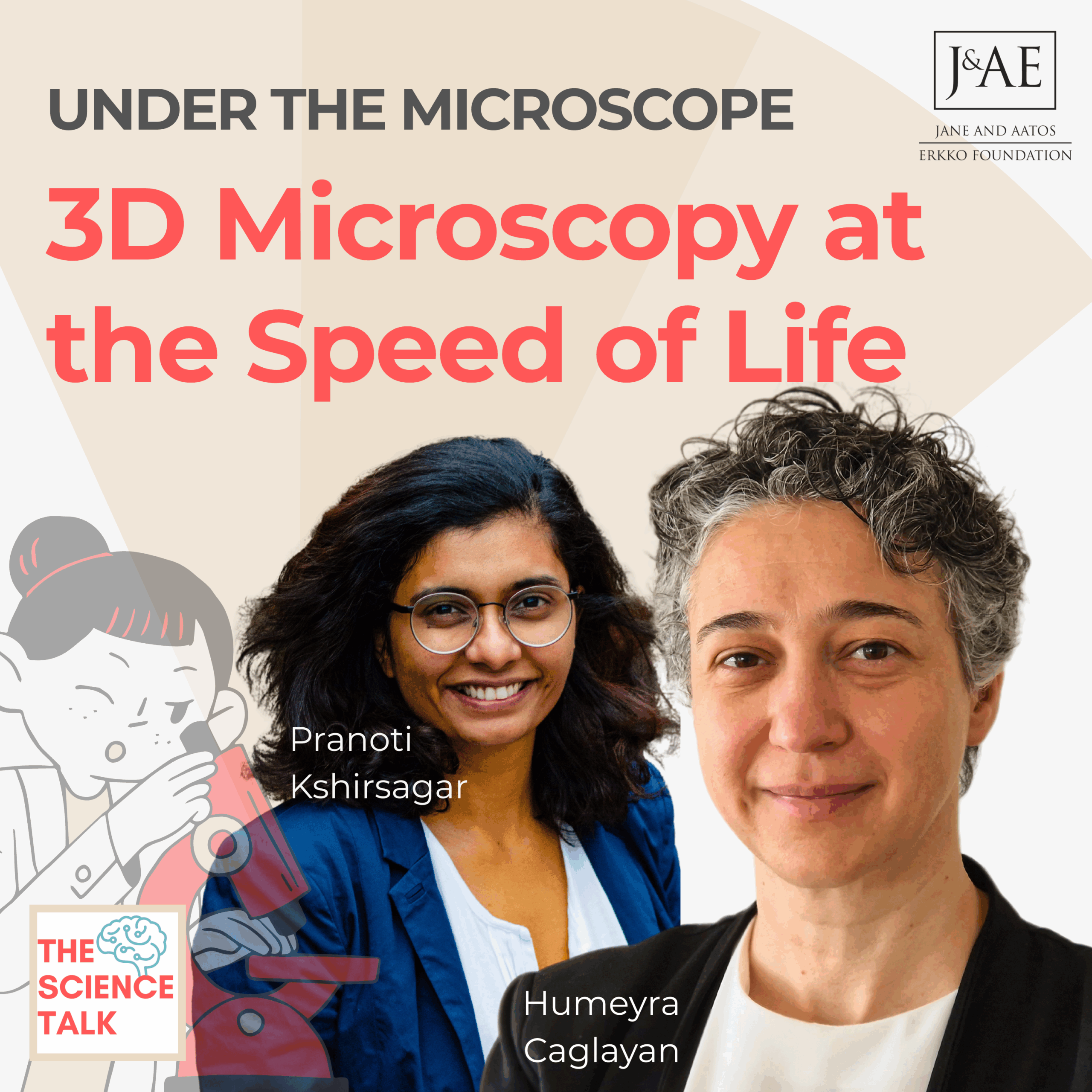
What if we could capture stunning 3D images of living cells, tissues—or even a tiny zebrafish brain—in real time, without staining, freezing, or harming them? Prof. Humeyra Caglayan’s research on snapshot 3D microscopy makes this possible.
In this episode
- What the 3D Microscopy project aims for: 3D images of living cells and organisms in a single snapshot, without killing the sample
- Why the team images zebrafish, which share about 70% of their genes with humans, and why brain activity needs faster images than one every 1.5 seconds
- How meta optics extend the depth of focus from a few microns to 300 to 500 microns, by mimicking the way we tilt our heads to see in 3D
- The team behind it at Tampere University: optics, image reconstruction software and biomedical partners
- Where the project stands after two years, and the next step: bringing the microscope to pathologists and operating rooms
Key Takeaways
🔬 Snapshot 3D Microscopy: A New Paradigm — Traditional imaging sacrifices speed for resolution in live biological systems. Snapshot 3D microscopy removes this trade-off, enabling high-resolution volumetric images in milliseconds.
🧠 Meta-Optics: Seeing Beyond the Surface — Novel meta-optics extend the depth of field, capturing entire biological volumes in focus without layer-by-layer scanning—faster, more accurate, and non-destructive.
⚡ Real-Time Imaging for Real-World Applications — From neuronal activity in zebrafish to surgical diagnostics, this technology opens new doors for both research and clinical use.
🤝 Interdisciplinary Innovation — Success here lies in collaboration between optical physicists, imaging software developers, and biomedical scientists.


Project Partners
- Prof. Humeyra Caglayan (Lead PI) — ORCID
- Haobijam Johnson Singh — Researcher Profile
- Jani Mäkinen — ORCID
- 3D Media Group — Research Group
- CIVIT — Research Infrastructure Unit
Further Reading
- “Flat lens for snapshot 3D microscopy” — Nanophotonics, 2023
- “A practical guide to scanning light-field microscopy with digital adaptive optics” — Nat Protoc 17, 2022
About the guest
Humeyra Caglayan is a Professor at Eindhoven University of Technology in the Netherlands and Tampere University in Finland. She leads the 3D Microscopy project, funded by the Jane and Aatos Erkko Foundation, which develops high-speed volumetric microscopy to image living cells in three dimensions.